Throat Cancer: Symptoms, Prevention Tips, and Early Detection

Throat cancer is a broad term encompassing various malignancies arising within the throat region. While the specific location may differ, all throat cancers share similar risk factors, symptoms, and early detection methods. This comprehensive guide delves deeper into the different types of throat cancer, explores the warning signs, and emphasizes the importance of preventive measures and early diagnosis.

Unveiling the Different Faces of Throat Cancer

The throat, a muscular passage connecting the mouth and nose to the windpipe (trachea) and esophagus, comprises several sub-regions. Cancers can develop in any of these areas, each with a distinct name:

  • Pharyngeal Cancer: This cancer affects the pharynx, a hollow tube extending from behind the nose to the top of the trachea.
  • Laryngeal Cancer: The larynx, also known as the voice box, is the target of this type.
  • Oropharyngeal Cancer: Located in the oropharynx (the middle part of the throat encompassing the back of the mouth, base of the tongue, and tonsils), this cancer can significantly impact swallowing and speech.
  • Nasopharyngeal Cancer: This cancer originates in the nasopharynx, the upper part of the throat situated behind the nose.
  • Hypopharyngeal Cancer: The hypopharynx, the bottom portion of the throat, is susceptible to this type of cancer.
  • Tonsil Cancer: As the name suggests, this cancer develops in the tonsils, the lymph tissue located on either side of the back of the throat.

Further classification exists within some of these categories based on the specific location within the sub-region. For example, glottic cancer affects the vocal cords, while supraglottic cancer involves the area above the vocal cords, and subglottic cancer targets the region below.

Symptoms of Throat Cancer

Early detection is crucial for successful throat cancer treatment. Here’s a breakdown of the common symptoms that warrant a visit to a healthcare professional:

  • Persistent Sore Throat: A sore throat that lingers for more than two weeks, even after trying home remedies, could be a red flag.
  • Hoarseness or Voice Changes: Changes in your voice, such as persistent hoarseness, raspy voice, or difficulty speaking clearly, can signal throat cancer.
  • Difficulty Swallowing (Dysphagia): Pain or a burning sensation while swallowing food or liquids could indicate throat cancer.
  • Lump in the Neck: A noticeable lump in the neck or throat area, especially if it’s painless and doesn’t go away, should be promptly evaluated by a doctor.
  • Ear Pain: Persistent pain in one ear, particularly if accompanied by other symptoms, can be a sign of throat cancer.
  • Chronic Cough: A cough that doesn’t subside despite treatment might be a cause for concern.
  • Unexplained Weight Loss: Losing weight unintentionally can be a symptom of various medical conditions, including throat cancer.
  • Breathing Difficulties: Difficulty breathing or noisy breathing can arise due to throat cancer obstructing the airway.
  • Bad Breath: Chronic bad breath that doesn’t improve with proper oral hygiene practices can be a symptom of throat cancer.
  • Fatigue: Unusual and persistent tiredness or fatigue can be a warning sign of throat cancer.

Seeking Medical Attention at Ayushman Hospital Dwarka:

If you experience any of these symptoms, it’s vital to schedule an appointment with an ear, nose, and throat (ENT) specialist at Ayushman Hospital & Health Services. We have a team of experienced doctors equipped with advanced diagnostic tools to accurately identify the cause of your symptoms and recommend the appropriate course of treatment. Early detection is critical for improving treatment outcomes and overall prognosis.

Preventive Measures for Throat Cancer

While there’s no guaranteed way to prevent throat cancer entirely, adopting certain lifestyle modifications can significantly reduce your risk:

  • Eliminate Tobacco Use: Tobacco use, including smoking and chewing tobacco, is the leading risk factor for throat cancer. Quitting smoking or avoiding it altogether is the single most impactful preventive step.
  • Limit Alcohol Consumption: Excessive alcohol consumption weakens the body’s defense mechanisms, increasing the risk of developing throat cancer. Moderating alcohol intake or abstaining from it altogether is recommended.
  • Embrace a Healthy Diet: A diet rich in fruits, vegetables, and whole grains provides essential vitamins, minerals, and antioxidants that may help protect against cancer.
  • Consider HPV Vaccination: The human papillomavirus (HPV) is linked to some types of throat cancer. Vaccination against HPV, particularly for adolescents and young adults, can offer protection.
  • Maintain Good Oral Hygiene: Regular dental checkups and proper oral hygiene practices can help maintain a healthy throat environment and potentially reduce the risk of throat cancer.
  • Minimizing Exposure to Harmful Substances: Limit exposure to harmful chemicals and pollutants, especially in occupational settings. If working with such substances is unavoidable, wear appropriate protective gear like masks and respirators.
  • Regular Medical Check-Ups: Schedule regular medical check-ups with your doctor. These check-ups may include a physical examination of the head and neck area, potentially including a throat scope to visualize the inside of your throat.

Conclusion

A throat cancer diagnosis can be overwhelming, but Ayushman Hospital & Health Services stands by your side throughout the journey. Our team of dedicated professionals provides comprehensive care, from diagnosis and treatment planning to supportive care and follow-up. With our expertise and advanced facilities, you can receive the best possible care for throat cancer and navigate the road to recovery with confidence.

Best Orthopaedic Surgeon in Dwarka

Experiencing bone and joint issues can be frustrating and limit your mobility. If you’re seeking expert care, look at the skilled orthopaedic doctors in Dwarka at Ayushman Hospital & Health Services. This blog post will introduce you to some of Dwarka’s leading orthopaedic surgeons in Dwarka and their areas of expertise.

At Ayushman, We Have The Best Orthopaedic Surgeon in Dwarka, Delhi

Ayushman Hospital & Health Services is a leading healthcare provider in Dwarka, offering comprehensive orthopaedic care. Our team of experienced orthopaedic doctors in Dwarka utilises advanced techniques and minimally invasive procedures whenever possible, leading to faster recovery times and reduced pain.

Our best orthopaedic doctors in Dwarka:

Dr. Raj Kumar:
Dr. Raj Kumar holds the position of Director and leads the Department of Orthopaedics and Joint Replacement at Ayushman Hospital. With over 30 years of experience, Dr. Kumar is a highly sought-after orthopaedic surgeon in Dwarka. His expertise lies in managing complex trauma cases, joint replacements (knee and hip), and spine surgeries. Dr. Kumar prioritizes minimally invasive techniques, ensuring a smoother recovery process for his patients.

Dr. Gaurav Khera:
Dr. Gaurav Khera is a Consultant Orthopaedic Surgeon in Dwarka at Ayushman Hospital. His area of focus encompasses a wide range of bone and joint disorders, with a particular interest in spine surgeries, trauma surgeries, and arthroplasty (joint replacement). Dr. Khera leverages his extensive training and experience to provide exceptional care for his patients in Dwarka.

Dr. Sourabh Shah:
Dr. Sourabh Shah brings his expertise in bone and joint disorders to Ayushman Hospital as a Consultant Orthopaedic Surgeon in Dwarka. He holds a special interest in total knee replacement and trauma management. Dr. Shah’s qualifications include graduating from Ruxmaniben Deepchand Medical College and undergoing specialist training at M.S. Ramaiah Medical College and Hospitals.


Dr. Avanish Kumar Srivastava:
Dr. Avanish Kumar Srivastava is a highly regarded Senior Consultant Orthopaedic Surgeon in Dwarka at Ayushman Hospital. His expertise encompasses various bone and joint conditions, with a focus on total hip and knee replacements. Dr. Srivastava’s educational background includes graduating from S.N. Medical College and undergoing advanced training at the Indian Spinal Injuries Centre.

How to Choose the Right Orthopaedic Surgeon in Dwarka?

Finding the right orthopaedic doctors in Dwarka is vital for receiving the best possible care. Here are some factors to consider when making your decision:

  • Area of Expertise: Orthopaedic doctors often specialize in specific areas, such as sports medicine, joint replacement, or trauma surgery. Consider your specific condition and choose a doctor with expertise in that area.
  • Experience: Experience is a valuable asset in any medical professional. Look for a doctor who has a proven track record of success in treating conditions similar to yours.
  • Patient Reviews: Reading online reviews from past patients can offer valuable insights into a doctor’s bedside manner, communication style, and overall patient experience.
  • Hospital Affiliation: Consider the facilities and amenities offered by the hospital where the doctor practices. Ayushman Hospital & Health Services is known for its advanced technology and commitment to patient comfort.

Conclusion

Ayushman Hospital & Health Services is a leading destination for orthopaedic care in Dwarka. The team of highly qualified orthopaedic doctors in Dwarka offers a comprehensive range of services, from diagnosis and treatment to rehabilitation. By considering the factors mentioned above, you can feel confident in selecting the right orthopaedic doctors in Dwarka to address your specific needs and get you back on the road to recovery.

Cancer Screening

Cancer screening is a proactive approach to healthcare, empowering you to take control of your well-being. It involves a series of tests designed to detect cancerous cells or precancerous abnormalities before they develop into full-blown cancer and cause symptoms. Early detection is crucial in the fight against cancer, significantly increasing the chances of successful treatment and improving overall survival rates.

Advantages of Early Detection

  • Enhanced Treatment Options: When cancer is caught in its early stages, it’s often confined to a smaller area, making it more susceptible to complete removal through surgery or targeted treatment with radiation or medication. Early diagnosis allows doctors to employ less aggressive and more effective treatment plans, minimizing side effects and improving the patient’s quality of life.
  • Improved Survival Rates: Studies have consistently shown a clear correlation between early detection and increased survival rates. Early-stage cancers often respond well to treatment, leading to a higher cure rate and a longer lifespan for patients.
  • Reduced Treatment Burden: Early intervention can significantly lessen the intensity and duration of treatment needed. This translates to a potential decrease in the financial burden associated with cancer care, as well as a reduction in the physical and emotional toll that extensive treatment can take on patients.
  • Minimizing Symptom Experience: Cancerous growths often cause a variety of unpleasant symptoms as they progress. Early detection allows for intervention before these symptoms manifest, sparing individuals from the physical and emotional distress associated with advanced cancer.

Why Early Detection is Worth It?

The fear of a positive test result can be a significant deterrent for some individuals when it comes to cancer screening. However, it’s crucial to understand that ignoring the possibility of cancer doesn’t make it disappear. Early detection is the key to effectively combating the disease and achieving a positive outcome.

A positive screening result can be unsettling, but it also empowers you to take immediate action. Early diagnosis allows for prompt treatment, potentially eradicating the cancer before it spreads and significantly improving your chances of a full recovery. Even if the result is negative, the peace of mind and reassurance gained from knowing your health status are invaluable.

A Spectrum of Screening Techniques

The landscape of cancer screening encompasses a variety of tests, each tailored to detect specific types of cancer. Here’s an overview of some commonly used methods:

Laboratory Tests:

  • Blood Tests: Certain blood tests can identify markers suggestive of cancer, although they may not be definitive for diagnosis.
  • Oral Cancer Screening Tests: These tests may involve visual inspection of the mouth and throat or the use of special rinses or lights to detect abnormalities.
  • Urine Tests: In some cases, urine tests can be used to screen for specific cancers, such as bladder cancer.
  • Tissue Samples: Biopsies involving the removal of a small sample of tissue for microscopic examination can definitively diagnose cancer.
  • Hormone Testing: Imbalances in hormone levels may be indicative of certain cancers, prompting further investigation.

Physical Exams and History Analysis:

A thorough physical examination often includes a visual inspection for lumps or other abnormalities. Additionally, your doctor will discuss your personal medical history and any relevant family history of cancer, which can help identify individuals at higher risk.

Genetic Testing: For individuals with a strong family history of certain cancers, genetic testing may be recommended to identify mutations that increase the risk. This allows for more targeted screening and early intervention.

Imaging Procedures: Imaging techniques like X-rays, mammograms, CT scans, and MRIs can create pictures of internal organs and tissues, potentially revealing suspicious abnormalities that warrant further investigation.

The Importance of Individualized Screening

The recommended screening tests and frequency of testing will vary depending on several factors, including your age, gender, family history, and overall health. It’s critical to discuss your risk profile with your doctor at Ayushman Hospital and Health Services to determine the most appropriate screening plan for you. Detecting cancer early is a potent weapon in the battle against this disease. By understanding the benefits of screening and overcoming the fear of a positive result, you can take charge of your health and potentially save your life.

Kidney Stones

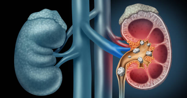
Kidney stones, those tiny but terrible masses of minerals, can wreak havoc on your urinary system. Affecting over 12% of the Indian population, they’re a common yet painful problem.

This blog dives deep into the world of kidney stones, exploring their causes, symptoms, diagnosis, and most importantly, prevention and treatment options.

Kidneys remove waste products from your blood, sending them out in your urine. But sometimes, minerals and salts in your urine can become concentrated, forming hard deposits – kidney stones. These stones can vary in size, from a speck to a bothersome pebble.

Causes of Kidney Stones

While the exact cause remains elusive, several factors can contribute to kidney stone formation:

  • Dehydration: Think of your urine as a solvent. When you don’t drink enough fluids, the urine becomes more concentrated, increasing the chance of minerals crystallizing and forming stones. Aim for plenty of water throughout the day!
  • Dietary Choices: A diet high in salt, sugar, and animal protein can tip the scales in favor of stone formation. Moderation is key!
  • Genetic Predisposition: If kidney stones run in your family, you might be more susceptible.
  • Underlying Medical Conditions: Certain medical conditions like gout, hyperparathyroidism, and Crohn’s disease can increase your risk.

Symptoms of Kidney Stones

Kidney stones often announce their presence with a bang – excruciating pain. Here are some red flags to watch out for:

  • Severe pain in your back, side, or groin: This pain can be sharp, stabbing, or come in waves, often radiating from your back to your lower abdomen.
  • Painful urination: Passing urine can become a burning sensation, accompanied by discomfort.
  • Difficulty passing urine: You might feel an urge to urinate frequently, but only manage to pass small amounts.
  • Blood in your urine: The stone could irritate the lining of your urinary tract, leading to blood in your urine.
  • Nausea and vomiting: The intense pain can sometimes trigger nausea and vomiting.

Diagnosis of Kidney Stones

If you experience these symptoms, consulting a doctor is crucial. Here’s what you might expect:

  • Urine test: This checks for blood, infection, or abnormal mineral levels.
  • Blood test: It helps assess your overall health and identify any underlying medical conditions.
  • Imaging tests: X-rays, CT scans, or ultrasounds can pinpoint the location and size of the stone.

Precautions for Kidney Stones

Here’s the good news: you can significantly reduce your risk of kidney stones with some lifestyle changes:

  • Hydration is Key: Aim for plenty of water throughout the day. Clear urine is a good indicator of adequate hydration.
  • Dietary Tweaks: Limit salt, sugar, and animal protein intake. Choose fruits, vegetables, and whole grains for a kidney-friendly diet.
  • Medication Management: Discuss any medications you take with your doctor, as some can increase your stone risk.
  • Manage Medical Conditions: If you have a condition linked to kidney stones, proper management is essential.

Treatment for Kidney Stones

The treatment for kidney stones depends on their size and location. Here are some common approaches:

  • Passing the Stone Naturally: For small stones, drinking plenty of fluids and pain medication might be enough to help you pass the stone.
  • Shockwave Lithotripsy: Sound waves are used to break down larger stones into smaller fragments that you can then pass.
  • Ureteroscopy: A thin, lighted tube is inserted through your urethra and bladder to reach the stone. Lasers or other tools can then be used to break down or remove the stone.
  • Percutaneous Nephrolithotomy (PCNL): For larger or complex stones, a small incision is made in your back, and a thin tube is inserted into your kidney to remove the stone.

Kidney Stone treatment at Ayushman Hospital and Health Services

Ayushman Hospital and Health Services stand as your partner in conquering kidney stones. We prioritize high-quality and affordable care. Our urology team is well-versed in diagnosing and treating stones using the latest techniques. Advanced diagnostic tools like X-rays, CT scans, and ultrasounds ensure accurate stone identification. Whenever possible, they prioritize minimally invasive procedures like ureteroscopy to minimize discomfort and recovery time. Additionally, we offer a holistic approach, including pain management and dietary counseling, to ensure a smooth recovery from your kidney stone troubles.

Frozen Shoulder

What is a Frozen Shoulder?

Frozen shoulder, also known as adhesive capsulitis, is characterized by stiffness and pain in the shoulder joint, significantly limiting movement. It can be debilitating, affecting daily activities like dressing, combing hair, or reaching overhead. While the exact cause of frozen shoulder remains elusive in some cases, understanding the different types, symptoms, diagnosis, and treatment options can help manage this condition effectively.

Understanding the Shoulder Joint

The shoulder joint is a ball-and-socket joint, where the ball (humeral head) of the upper arm bone fits into the socket (glenoid) of the shoulder blade. A capsule of connective tissue surrounds the joint, providing stability and allowing for a wide range of motion.

What Causes Frozen Shoulder?

There are two main categories of frozen shoulder:

  • Primary Frozen Shoulder (Idiopathic Adhesive Capsulitis): This occurs without a readily identifiable cause. It is more common in people with diabetes, thyroid problems, and those aged 50-60, particularly women.

  • Secondary Frozen Shoulder: This develops due to another condition or event that restricts shoulder movement for an extended period. Examples include shoulder surgery, fracture healing, stroke, or even heart surgery.

Symptoms of Frozen Shoulder

The symptoms of a frozen shoulder typically progress through three stages:

  • Freezing Stage (2-9 Months): This stage is characterized by increasing pain, often worse at night and with rest. The shoulder joint becomes progressively stiffer, and muscle spasms can occur.

  • Frozen Stage (4-12 Months): Pain intensity gradually decreases in this stage. However, the stiffness becomes more prominent, significantly restricting both active (self-initiated) and passive (assisted) movement of the shoulder.

  • Thawing Stage (5-24 Months): The pain subsides further in this stage, and the range of motion slowly improves. This stage can be quite unpredictable, with some individuals regaining full mobility within 6 months, while others may experience residual stiffness even after 3 years.

How is Frozen Shoulder Diagnosed?

Diagnosis of a frozen shoulder is primarily based on a thorough physical examination by a doctor who will assess your range of motion and pain levels. Imaging tests like X-rays are usually not required for diagnosis but may be helpful to rule out other conditions mimicking frozen shoulder symptoms.

  • Ultrasonography: This is a quick and easy way to visualize the shoulder joint and identify the thickening of the ligaments or adhesions. However, the accuracy depends on the operator’s skill.

  • MRI: While more expensive than ultrasound, MRI offers a more detailed picture of the joint and surrounding tissues. It can help identify loss of the axillary recess, a hallmark of adhesive capsulitis.

How is Frozen Shoulder Treated?

The primary goals of frozen shoulder treatment are pain management and improvement of shoulder mobility. The specific treatment approach depends on the type and stage of the frozen shoulder.

Treatment for Secondary Frozen Shoulder

Secondary frozen shoulder is generally easier to manage than primary frozen shoulder. The mainstay of treatment involves:

  • Physical Therapy: A physical therapist will guide you through exercises designed to improve your range of motion and flexibility. These exercises can be performed independently at home or under the supervision of a therapist. Initially, the focus might be on gentle range-of-motion exercises followed by gradual progression to more challenging exercises without causing strain.

  • Moist Heat Therapy: Applying moist heat packs to the shoulder can help relax muscles and improve blood flow, promoting pain relief.

  • Non-Steroidal Anti-Inflammatory Drugs (NSAIDs): Over-the-counter pain relievers like ibuprofen or naproxen can help manage pain and inflammation.

Treatment for Primary Frozen Shoulder

The treatment approach for primary frozen shoulder varies depending on the stage of the condition:

  • Freezing Stage:

    • Pain Management and Physical Therapy: This is the first line of treatment. Pain relievers like NSAIDs can help manage pain, while physical therapy exercises can help maintain some degree of movement and prevent further stiffness.

    • Intra-articular Steroid Injections: Injections of corticosteroids directly into the shoulder joint can be beneficial for managing severe pain.

  • Frozen Stage:

    • Physical Therapy: Here, the focus of physical therapy shifts towards joint mobilization techniques to break down adhesions and improve range of motion.

    • Intra-articular Steroid Injections: Generally not recommended in this stage as they offer minimal benefit.

    • Hydro-distension: This procedure involves injecting a large volume of saline solution into the joint to stretch the capsule and break down adhesions. While promising in theory, it has not yielded consistent results in clinical practice.

    • Manipulation Under Anesthesia (MUA): This controversial procedure involves manipulating the shoulder joint under anesthesia to force movement and break down adhesions. However, it carries a risk of complications like tendon tears, ligament ruptures, and even fractures.
  • Arthroscopic Capsular Release: This minimally invasive surgical procedure is considered the treatment of choice for patients who fail to improve with physiotherapy after 3 months. A surgeon uses a small camera and specialized instruments inserted through tiny incisions to cut or release the tightened capsule, restoring shoulder movement. Studies have shown this procedure to be highly effective with good patient satisfaction.

Secondary Prevention of Frozen Shoulder

Following shoulder surgery, consistent physical therapy is crucial to maintain joint mobility and prevent stiffness. Recovery time varies depending on the individual and the type of surgery, typically ranging from 6 weeks to 3 months.

Here are some additional points for preventing a recurrence of a frozen shoulder:

  • Address Underlying Conditions: If you have diabetes or other risk factors, managing those conditions can help reduce the risk of developing a frozen shoulder again.

Avoid Prolonged Immobilization: After an injury or surgery, it’s important to follow your doctor’s recommendations for gentle movement exercises to prevent stiffness from developing.

What is a Complete Heart Block

Heart block, also known as atrioventricular (AV) block, is a problem with how the heart’s electrical system works. When someone has a heart block, their heartbeat becomes irregular and slow. Sometimes, it might even pause for around 20 seconds at a time. This happens because something is blocking or slowing down the electrical signals that control the heart’s rhythm. This blockage can occur because of damage or injury to the heart valves or muscles.

Because the heart itself is the one causing the blockage, it usually doesn’t require direct treatment. However, the underlying health issues that led to the heart block might need attention.

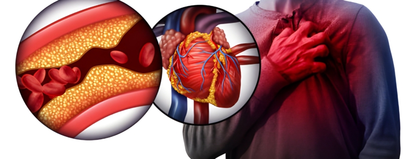
Heart block can cause symptoms like feeling your heart beating irregularly (palpitations), fainting, feeling dizzy, or even chest pain. Depending on how severe it is, heart block can be dangerous. For example, a complete heart block, also called third-degree heart block, can make existing health problems worse. It could even lead to passing out or sudden cardiac arrest.

What is a Heart Block?

Heart block messes with how the heart’s electrical signals work. Normally, your heart beats around 60 to 100 times per minute when you’re resting. Each beat pushes blood through your body. Your heart’s muscle contractions are controlled by electrical signals that move from the upper chambers (atria) to the lower chambers (ventricles).

Partial heart block happens when these electrical signals are slowed down or partly blocked. This can mess up the timing of your heartbeats and how well your heart pumps blood.

A complete heart block is when the electrical signals are completely messed up or stopped. This can slow your heartbeat down to around 40 times per minute. Even tiny changes in these signals, lasting just a fraction of a second, can cause heart block.

Sometimes, heart block makes it hard for your heart to pump blood around your body. This means less oxygen gets to your muscles and organs, including your brain, which can mess with how well they work.

Types of Heart Block

Heart block comes in three types:

First-degree heart block: This is a minor hiccup in the heartbeat that might make you feel like your heart skips a beat. It’s usually not a big deal and often doesn’t need treatment.

Second-degree heart block: In this type, some electrical signals struggle to get through to your heart. This can lead to skipped or dropped heartbeats. When the signal from the upper chambers (atria) can’t make it to the lower chambers (ventricles), the heart might not pump properly. It can make you feel dizzy, and sometimes you might need a pacemaker to help regulate your heartbeat.

Complete heart block or third-degree heart block: This happens when the electrical signals can’t travel properly between the upper and lower chambers of the heart. It’s pretty common among people with heart issues. It can be caused by surgery damaging the atrioventricular node (AV node), or it might happen on its own. To fix the heart’s rhythm, you’d need an artificial pacemaker. Without one, there’s a higher risk of a heart attack for people with complete heart block.

Heart Block Symptoms

When someone has complete heart block, they might experience:

  • Chest pain
  • Feeling like their heart is fluttering, pounding, or skipping (palpitations)
  • Dizziness or feeling like they might faint (pre-syncope)
  • Shortness of breath
  • Extreme tiredness or fatigue
  • Fainting (syncope)
  • Slow heartbeat (bradycardia)

These symptoms can be serious and even life-threatening. If you or someone else has these symptoms, it’s important to call for an ambulance or contact your local emergency medical service right away.

Causes of Heart Block

Most people diagnosed with complete heart block already have an underlying heart condition, such as congenital heart disease, cardiomyopathy, or coronary heart disease. Age can also play a role, as the heart’s electrical pathways age too. Certain medications and imbalances in electrolytes can also contribute to complete heart block.

Diagnosing Heart Block

To diagnose heart block, a person would typically visit a heart specialist called a cardiologist for a complete heart evaluation. The cardiologist would review the person’s medical history, including any past heart tests they’ve had.

The doctor might ask questions about the person’s overall health, diet, activity level, and family medical history. They’ll also ask about any medications, including over-the-counter and prescription drugs, as well as alcohol, drug use, or smoking habits.

Next, the person will undergo a thorough physical exam. The doctor will check their pulse, measure their heart rate and rhythm, and look for signs of heart failure, like fluid retention in the feet or legs.

Heart block is diagnosed using an Electrocardiogram (ECG or EKG), which records the heart’s electrical activity. This test creates a graph showing the heart’s rhythm, rate, and timing of electrical signals as they move through the heart.

If the doctor needs to monitor the person’s heart over a longer period, they might recommend wearing a Holter Monitor. This is a small, portable ECG machine that continuously monitors the heart for 24 to 72 hours. It can help detect problems that may not show up on a standard ECG done at rest.

Treatment of  Heart Block

Complete heart block can be very serious, so the specialist might suggest medication or implanting a pacemaker, depending on how severe it is.

Medications: The patient might get prescribed anti-arrhythmic medications. These drugs can change the heart’s electrical signals and help prevent disruptions in its electrical system.

Pacemaker: Implanting a pacemaker involves a small surgery. The device has two wires that attach to the right side of the heart. It acts like a backup electrical system, reminding the heart to beat at a normal rate if it slows down or stops.

Complete heart block is a big deal, so getting the right diagnosis and treatment is super important. Make sure to see a highly skilled and experienced heart specialist for proper care.

Moyamoya Disease

Moyamoya disease is a rare but serious cerebrovascular condition. It affects the blood vessels supplying blood to the brain. This article explores Moyamoya disease, its causes, symptoms, diagnosis, treatment options, and living with the condition.

What is Moyamoya Disease?

Moyamoya, meaning “puff of smoke” in Japanese, refers to the abnormal tangled network of small blood vessels that develop at the base of the brain in this disease. This compensatory network forms in response to the gradual narrowing of the major arteries supplying blood to the brain.

The exact cause of Moyamoya disease remains unknown. However, several factors are believed to contribute to its development:

  • Genetics: Genetic mutations might play a role, especially those related to blood vessel development.
  • Ethnicity and Geography: The disease is more prevalent in people of Asian descent, particularly in East Asian countries.
  • Environmental Factors: Environmental triggers might influence the disease, but further research is needed.

Symptoms and Stages of Moyamoya Disease

Symptoms of Moyamoya disease can vary depending on the individual and the stage of the disease. They may appear suddenly or develop gradually over time. Here’s a breakdown of potential signs:

  • Transient Ischemic Attacks (TIAs): Brief episodes of neurological dysfunction resembling mini-strokes, causing temporary weakness, numbness, or speech difficulties.
  • Ischemic Strokes: Sudden onset of weakness, paralysis, or sensory problems on one side of the body, difficulty speaking, or loss of consciousness due to reduced blood flow.
  • Seizures: Uncontrolled electrical activity in the brain, leading to seizures in some individuals.
  • Headaches: Frequent headaches, ranging from mild to severe.
  • Cognitive Changes: Issues with memory, thinking, and other cognitive functions, particularly in children.
  • Vision Problems: Blurred vision, vision loss in one eye, or other visual disturbances.
  • Weakness or Paralysis: Sudden weakness or paralysis on one side of the body (hemiparesis/hemiplegia) during strokes.

Moyamoya disease typically progresses through stages:

  1. Early Stage (Pre-Stroke): No significant symptoms or subtle, easily overlooked ones. The hallmark is the development of “puff of smoke” collateral vessels.
  2. Symptomatic Stage (TIA): Individuals experience symptoms like TIAs due to reduced blood flow.
  3. Stroke Stage: Increased risk of strokes with lasting neurological deficits.
  4. Late Stage (Late-Onset Stroke): Higher risk of recurrent and severe strokes with significant neurological impairment.

Progression rates vary, and not everyone experiences all stages. Early diagnosis and intervention are crucial to potentially slow or halt disease progression and reduce complications.

Diagnosis of Moyamoya Disease

Diagnosing Moyamoya disease involves a combination of assessments:

  • Medical History and Physical Examination: The doctor reviews your symptoms, medical history, and performs a physical exam to assess neurological function.
  • Imaging Tests: These are vital for confirmation:
    • Magnetic Resonance Imaging (MRI): Provides detailed brain images to detect abnormalities in blood vessels and signs of reduced blood flow or stroke.
    • Magnetic Resonance Angiography (MRA): This specialized MRI technique focuses on blood vessels to visualize changes in the brain’s arteries.
    • CT Scan: This may be used to identify signs of stroke or brain abnormalities.
    • Cerebral Angiography: An invasive test involving contrast dye injection to directly visualize the characteristic blood vessel changes of Moyamoya disease.
  • Other Tests: Blood tests to rule out other conditions and cognitive function tests might be recommended.

The definitive diagnosis often relies on the characteristic appearance of narrowed blood vessels and the abnormal collateral network seen in imaging studies.

Treatment Options for Moyamoya Disease

Treatment aims to improve blood flow to the brain, prevent strokes, and manage symptoms. The primary approach is:

Surgical Revascularization:

  • Direct Bypass Surgery: Connects a healthy scalp blood vessel to a brain surface blood vessel, creating a new blood flow route.
  • Indirect Bypass Surgery: Places surgical material on the brain surface to stimulate the growth of new blood vessels over time, enhancing blood flow.

Medication Management:

  • Antiplatelet Agents: Reduce blood clot risk to prevent strokes.
  • Blood Pressure Medication: Controls hypertension to manage the disease and minimize further complications.
  • Anticonvulsants: Manage and prevent seizures if they occur.

Ongoing Monitoring and Care: 

  • Regular check-ups with a specialist are essential to monitor disease progression, assess treatment effectiveness, and identify any emerging complications.

Living with Moyamoya Disease

While living with Moyamoya disease has challenges, proper management allows individuals to lead fulfilling lives. Here are some key considerations:

Lifestyle Adjustments

  • Healthy Lifestyle: A balanced diet, regular exercise, and avoiding smoking positively impact overall health.
  • Stress Management: Techniques like relaxation or counseling can be beneficial as stress may worsen symptoms.

Emotional and Social Support

  • Support Networks: Connecting with support groups or others with Moyamoya disease provides emotional support and a sense of community.
  • Mental Health Support: Seeking professional help can be beneficial, especially during difficult times.

Daily Living

  • Adaptive Strategies: Individuals might need to adapt activities or use assistive devices if symptoms affect daily life.
  • Career and Education: Adjustments in workload or learning accommodations may be necessary to balance work/education with disease management.
  • Educating Others: Raising awareness among family, friends, and the broader community fosters understanding and support.

Planning for the Future

  • Advanced Directives: Discussing future medical care preferences with family and healthcare providers ensures your wishes are respected.
  • Financial Planning: Considering long-term medical care and possible lifestyle changes promotes financial preparedness.

Conclusion

Moyamoya disease is a complex condition, but with proper diagnosis, treatment, and management, individuals can live fulfilling lives. Early diagnosis is crucial, and consulting a specialist at a renowned institution experienced in Moyamoya disease is vital for optimal care.

Top Five Mistakes After Knee Replacement Surgery

Regaining the ability to move easily and free from chronic knee pain is a life-changing prospect. Knee replacement surgery offers just that, with a success rate of 90-95% of total knee replacements functioning well even after ten years.

However, achieving this optimal outcome hinges on a crucial factor – your active participation in the recovery process. Like scaling a mountain, successful recovery from knee replacement surgery requires a well-defined plan, perseverance, and the right tools. This comprehensive guide equips you with the knowledge and strategies to navigate this journey effectively.

Understanding Your Terrain: Pre-Surgery Preparation

Even before surgery, proactive steps can pave the way for a smoother recovery. Here’s what you can do:

  • Education is Power: Familiarize yourself with the surgical procedure, potential risks and benefits, and the recovery timeline. Discuss any anxieties you have with your doctor.
  • Building Your Support System: Identify individuals who can assist you with daily tasks like cooking, grocery shopping, and transportation during the initial recovery phase.
  • Optimizing Your Fitness: If possible, engage in pre-habilitation exercises to strengthen the muscles surrounding your knee joint. This improves your baseline strength and prepares your body for post-surgical rehabilitation.
  • Conquering the Clutter: Declutter your living space to minimize obstacles that could lead to falls. Ensure grab bars are installed in your bathroom, and remove any loose rugs that could cause slips.

The Postoperative Plan

Your doctor will provide you with a detailed postoperative plan, and your roadmap to recovery. This plan typically includes:

  • Pain Management: Medications will be prescribed to manage pain and inflammation, allowing your body to focus on healing. Open communication with your doctor is crucial to address any concerns about side effects or pain control.
  • Wound Care: Maintaining a clean and properly dressed surgical site is essential to prevent infections. Your doctor will provide specific instructions on wound care.
  • Range of Motion Exercises: Early and gentle exercises will be initiated to prevent stiffness and promote flexibility in your new knee joint.
  • Weight-Bearing Restrictions: Your doctor will advise you on the appropriate amount of weight to bear on your knee initially. This may involve using assistive devices like walkers or crutches for some time.
  • Physical Therapy: Formal physical therapy sessions are a cornerstone of recovery. A dedicated therapist will guide you through targeted exercises to strengthen the muscles around your knee, improve flexibility, and restore your range of motion.

5 Common Mistakes After Knee Replacement Surgery

1. Neglecting the Postoperative Plan:

Adhering to your prescribed postoperative regimen and doctor’s advice is vital. Failing to do so can cause complications and impede your recovery progress. For instance, inadequate wound care and bandaging may increase the risk of infection, hindering your healing process. Thus, it’s imperative to stick to your plan diligently, attend all follow-up appointments, and promptly address any unusual symptoms or swelling by consulting your doctor.

2. Inadequate Medication Management:

Proper medication intake is crucial for managing post-surgery pain and inflammation. Skipping doses due to concerns about addiction or side effects can prolong discomfort and delay healing. Openly discussing any concerns or adjustments with your healthcare provider ensures effective pain management while minimizing risks.

3. Rushing Rehabilitation or Returning to Work Prematurely:

Although the desire to resume normal activities promptly is understandable, pushing yourself too hard can compromise your recovery. Overexertion strains the newly replaced knee joint and may lead to complications. Embracing patience and allowing your body the time it needs to heal is essential for optimal outcomes.

4. Neglecting Nutrition:

Nutrition plays a pivotal role in tissue repair and overall well-being during recovery. A balanced diet rich in essential nutrients accelerates healing. Prioritizing fruits, vegetables, lean proteins, and whole grains supports your recovery journey and enhances your overall health.

5. Underestimating the Importance of Physical Therapy:

Physical therapy is indispensable for knee replacement recovery, aiding in mobility, flexibility, and range of motion. Skipping or insufficiently participating in recommended sessions may impede progress and result in stiffness. Regular attendance and active engagement in physiotherapy sessions are critical for achieving optimal outcomes. Collaborating with your physical therapist to maintain motivation and interest in exercises ensures a successful rehabilitation journey.

Conclusion

Knee replacement surgery offers the potential for a renewed lease on life, free from chronic knee pain. By following the strategies outlined in this guide, actively participating in your recovery process, and embracing a commitment to healthy habits, you can conquer the climb and reach the summit of a strong, healthy knee. Remember, you are not alone on this journey. Your doctor, physical therapist, and support system are there to guide and encourage you every step of the way. Embrace the challenge, celebrate your progress, and look forward to a future filled with renewed mobility and freedom.

Kidney Infection Treatment in Delhi

Your kidneys, bean-shaped organs that filter waste from your blood, can become infected. This infection, called pyelonephritis, usually starts in the bladder or lower urinary tract and travels up to the kidneys. Bacteria are the main culprits, but sometimes viruses can be to blame.

Kidney Infection Symptoms

Kidney infections develop quickly, often within hours or a day. Here’s what to watch for:

  • Pain: This can be in your lower back or side, and sometimes even in your groin.
  • Urinary Issues: You might feel pain or burning when you pee, and you might need to go more often than usual, even feeling an urgent need to go right after emptying your bladder.
  • Fever and Chills: These are common signs your body is fighting an infection.
  • Other Symptoms: You might also experience nausea, vomiting, loss of appetite, or feel generally weak and unwell.

Remember: Kidney infections often start as a urinary tract infection (UTI). If you have a UTI, you might also notice:

  • Cloudy or bloody urine
  • Urine with a strong smell
  • Difficulty emptying your bladder completely

Don’t Ignore Confusion: In some cases, especially with older adults, a kidney infection might confuse a symptom.

Early diagnosis and kidney infection treatment in Delhi are crucial. If you experience any of these symptoms, see a doctor right away.

Causes of Kidney Infection

While viruses can rarely cause kidney infections in healthy individuals, bacteria are the usual culprits. These tiny invaders typically enter the urethra and travel up the urinary tract. This upward journey allows them to reach the kidneys and potentially cause an infection.

Some common troublemakers include:

  • E. coli: This infamous bacteria is a frequent offender, often residing in your intestines before making a wrong turn.
  • Enterobacter, Staphylococcus, and Proteus mirabilis: These are just a few other bacterial hitchhikers that can take advantage of a weakened urinary tract defense system.

Diagnosing a Kidney Infection

Doctors use a combination of approaches to diagnose a kidney infection. Here’s what to expect:

  • Doctor’s Checkup: First, the doctor will assess your overall health, including temperature, blood pressure, and any back pain or tenderness.
  • Medical History: Discussing your symptoms and any recent UTIs can help identify risk factors.

Tests to Confirm the Culprit

  • Urine Test (Urinalysis): This is a key test to check for white blood cells, red blood cells, and bacteria in your urine.
  • Blood Tests: These can reveal infection markers and potential kidney function issues.

Imaging for a Closer Look (not always needed)

  • Ultrasound: This painless test uses sound waves to create images of your kidneys and bladder to check for abnormalities.
  • CT Scan or MRI: In some cases, more detailed pictures from a CT scan or MRI might be needed.

Additional Exams (depending on gender):

  • Men: A rectal exam can assess the prostate for issues affecting urine flow.
  • Women: A pelvic exam may be performed to rule out pelvic inflammatory disease.

Kidney Infection Treatment in Delhi

The good news: most kidney infections can be successfully treated! The approach depends on your case and how severe the infection is. Here’s what to expect:

  • Antibiotics are Your Ally in Kidney Infection Treatment: This is the first line of defense. Your doctor will prescribe a specific antibiotic based on the bacteria causing the infection and your medical history. Be sure to take the full course, even if you start feeling better, to completely knock out the infection.
  • Hydration is Key: Drinking plenty of fluids helps flush out bacteria from your urinary tract. Aim for clear liquids like water, cranberry juice (unsweetened is best), and clear broth.
  • Pain Relief on Deck: Over-the-counter pain relievers like acetaminophen can help manage discomfort.
  • Hospital Care for Tough Cases: If your infection is severe, you might need hospitalization. Here, you’ll receive intravenous (IV) fluids and antibiotics directly into your bloodstream to fight the infection more aggressively.
  • Surgery in Special Cases: If a blockage like an enlarged prostate or kidney stone is contributing to your infection, surgery might be needed to remove the obstruction and prevent future problems.

Risk Factors for Kidney Infection

While anyone can get a kidney infection, certain factors can increase your risk:

  • Plumbing Problems: Women have a shorter urethra, making it easier for bacteria to enter the urinary tract. Pregnancy can also put pressure on the bladder, increasing risk.
  • Blockages Down the Line: Kidney stones, enlarged prostate in men, or a prolapsed uterus can all hinder urine flow, allowing bacteria to multiply.
  • Weakened Defenses: Conditions like HIV or diabetes and medications that suppress the immune system can make you more prone to infections, including kidney infections.
  • Backflow Issue: A condition called vesicoureteral reflux (VUR) causes urine to flow backward from the bladder to the kidneys, increasing infection risk.

By understanding these risk factors, you can be more aware of potential problems and seek medical attention promptly if symptoms arise.

Potential Complications of Kidney Infections

Kidney infections are treatable, but neglecting them can lead to serious problems:

  • Kidney Abscess: If left untreated, the infection can create a pus-filled pocket within your kidney. This can cause severe pain, blood in your urine, and weight loss.
  • Sepsis: This life-threatening condition occurs when the infection spreads throughout your body, causing a dramatic drop in blood pressure. It can lead to organ failure, including kidney failure, and even death. Symptoms include difficulty breathing, seizures, and coma.
  • Blood Clots and Chronic Kidney Disease: A kidney infection can increase your risk of blood clots in the veins around your kidneys. These clots can damage kidney tissue and potentially lead to chronic kidney disease.
  • Acute Kidney Failure: In severe cases, an untreated kidney infection can cause your kidneys to shut down completely. This condition, called acute kidney failure, requires dialysis to remove waste products from your blood.

Early diagnosis and kidney infection treatment in Delhi are essential to prevent these complications. If you experience any symptoms of a kidney infection, see a doctor right away.

Prevention of Kidney Infection

The good news is that many kidney infections can be prevented! Here are some simple steps you can take to reduce your risk:

  • Hydrate, Hydrate, Hydrate! Drinking plenty of fluids, especially water, helps flush out bacteria from your urinary tract. Aim for eight glasses a day or more.
  • Fiber is Your Friend: A diet rich in fiber keeps your digestive system moving smoothly, which can help prevent constipation and reduce your risk of UTIs that might travel to your kidneys.
  • Go When You Gotta Go: Don’t hold back on urination. Holding urine allows bacteria to multiply in your bladder.
  • Post-Intercourse Power Flush: Empty your bladder soon after sexual intercourse to help flush out any bacteria that may have been introduced.
  • Keep it Clean: Practice good hygiene by washing your genital area with plain water daily. Avoid harsh soaps or douches that can irritate the delicate skin in this area.
  • Wipe Front to Back: This helps prevent bacteria from spreading from your anus to your urethra.

By following these tips, you can take charge of your kidney health and reduce your chances of getting a kidney infection.

When to See a Doctor for Kidney Infection Treatment in Delhi

Kidney infections can worsen quickly. Here’s when to seek medical attention:

  • Pain in Your Back or Side: A dull ache or sharp pain in your lower back or side, near your ribs, could be a sign of a kidney infection.
  • Fever and Chills: A high fever (often above 101°F or 38.3°C) accompanied by chills is a red flag.
  • Burning When You Pee: Pain or a burning sensation while urinating is a common symptom.
  • Frequent Urination: Having to urinate more often than usual, even in small amounts, can be a sign of infection.
  • Urgent Urination: A sudden urge to urinate, even if you just went, could be a problem.
  • Blood in Your Urine: Seeing blood in your urine is a cause for concern and requires a doctor’s evaluation.
  • Feeling Gross: Generally feeling unwell, weak, nauseated, or vomiting can all be signs of a kidney infection.

Conclusion

Kidney infections can affect individuals of any gender and often progress swiftly. While many cases can clear up with sufficient hydration and antibiotics, others may necessitate further intervention. Maintaining proper hygiene and self-care can help prevent kidney infections altogether.

If you have any concerns about kidney infections or suspect you might have one, please consult a nephrologist at Ayushman Hospital & Health Services for proper diagnosis and treatment.